Your Exam

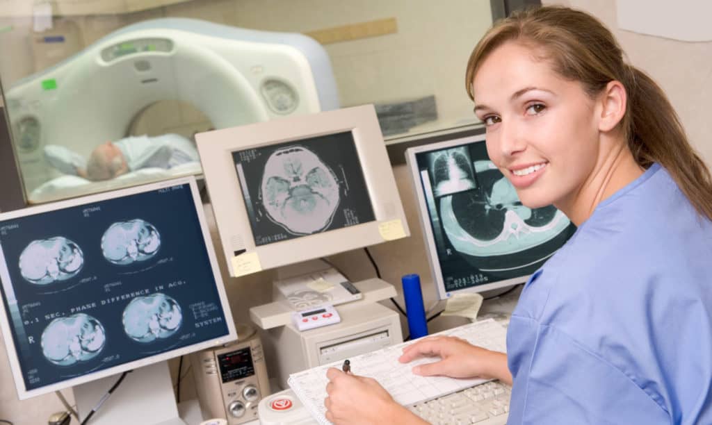
Computerized Axial Tomography Scan (CT or CAT Scan)
A CT Scan is a procedure that combines many x-ray images with the aid of a computer to generate cross-sectional and three-dimensional views of anatomy. CT Scans are performed to analyze the internal structures of various parts of the body. CT Scans can identify normal and abnormal structures and be used to guide procedures.
CT Scans are used to examine many areas of the body including brain, sinus, neck, chest, spine, abdomen, pelvis, and extremities. CT Scans can also be beneficial for evaluating the heart and lungs.
Some CT Scans require contrast material to be injected intravenously or administered by mouth in order to increase the distinction between various organs or areas of the body that have circulation.
CT Scans are very low-risk procedures. The amount of radiation the patient receives during a CT Scan is minimal. CT Scans provide a clear, accurate display of tissues without superimposition of structures. CT Scans may detect disease processes and lesions at an earlier stage than with conventional X-rays. CT Scans provide a wide range of imaging and the technique does not have to be limited to specific organs.
Learn more about the importance of early detection through CT lung cancer screening.
Magnetic Resonance Imaging (MRI)
MRI uses radio frequency waves and a strong magnetic field rather than X-rays to provide detailed images of the body. MRI is an invaluable tool in early diagnosis.
It assists physicians in evaluating the function as well as the structure of many organs. MRI is useful for the diagnosis of many conditions, including stroke, cancer, heart and vascular disease, as well as joint and musculoskeletal disorders. MRI evaluates body structures that may not be as visible with other imaging methods.
MRI is an alternative to traditional x-ray based and ultrasound imaging techniques. Breast MRI and MRI-guided breast biopsy have proven beneficial for early diagnosis of breast cancer and is many times used in addition to mammography
MRI is an emerging tool for early diagnosis of prostate cancer. The benefit of this new technology for patients is that the MR images taken can potentially identify specific areas within the gland that are suspicious and require additional evaluation
MRI requires specialized equipment and trained physicians to interpret the exam.
Nuclear Medicine
Nuclear medicine is a branch of medical imaging that uses small amounts of radioactive material to diagnose and determine the severity of or treat a variety of diseases. These diseases include cancer, diseases of the gastrointestinal tract, diseases of the bones, and diseases of the endocrine system. Because nuclear medicine procedures are able to evaluate molecular activity within the body, they offer the potential to identify disease in its earliest stages and to evaluate a patient’s response to therapeutic interventions.
Nuclear medicine imaging procedures are noninvasive and, with the exception of injections, are usually painless. These imaging scans use radioactive materials called radiopharmaceuticals or radiotracers.
Depending on the type of nuclear medicine exam, the radiotracer is either injected into the body, swallowed or inhaled as a gas and eventually accumulates in the organ or area of the body being examined. Radioactive emissions from the radiotracer are detected by a special camera or imaging device that produces pictures and provides molecular information.
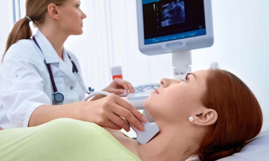
Ultrasound
Diagnostic Ultrasound, also called ultrasound scanning or sonography, produces pictures of the inside of the body using sound waves. It involves the use of a small transducer (probe) and ultrasound gel placed directly on the skin. High-frequency sound waves are transmitted from the probe through the gel into the body. The transducer collects the sounds that bounce back and a computer then uses those sound waves to create an image.
Ultrasound examinations do not use ionizing radiation (as used in x-rays), thus there is no radiation exposure to the patient. Because ultrasound images are captured in real-time, they can show the structure and movement of the body’s internal organs, as well as blood flowing through blood vessels, enabling medical professionals to use both sounds and visuals to assess a patient’s health.
Positron Emission Tomography (PET CT)
Positron Emission Tomography (PET) technology, often used in combination with CT imaging, uses a scanner and a radiopharmaceutical, which is injected into a patient’s vein to make detailed, computerized pictures of areas inside the body. A PET-CT scan consists of 2 parts: first, a CT scan to pinpoint the location for the PET, and then the PET scan itself. During a PET scan, a ring of detectors picks up radiation signals from the patient’s body coming from previously injected radiopharmaceuticals. The computer then analyzes the information and constructs an image of the targeted area.
X-ray/Fluoroscopy
X-RAY technology lets doctors see straight through human tissue to examine bones.
Fluoroscopy is a study of moving body structures–similar to an X-ray “movie.” A continuous X-ray beam is passed through the body part being examined. The beam is transmitted to a TV-like monitor so that the body part and its motion can be seen in detail.
Bone Density Scan
Bone density scanning, also called dual-energy x-ray absorptiometry (Dexa) or bone densitometry, is a specialized form of x-ray technology that is used to measure bone loss. Dexa is today’s established standard for measuring bone mineral density (BMD)
Mercy Breast Center
2D and 3D Mammography (Tomosynthesis)
The Mercy Breast Center offers both traditional 2D mammography and 3D Tomosynthesis. While most patients won’t notice a difference between the two, 2D mammography takes traditional images from the top, bottom, and side, while 3D mammography creates slices of images, like a CT scanner. Some women may benefit from a 3D mammogram, including women with dense breasts. Because of the slices, 3D mammography can produce clearer images, which can reduce stressful callbacks for repeat Talk with your doctor about whether a 2D or 3D mammogram is most appropriate for you.
Image-Guided Breast Biopsy
If your mammogram shows an area of concern, we offer ultrasound-guided, MRI guided, or stereotacticly-guided breast biopsy. Using these advanced imaging techniques allows your radiologist to accurately view your abnormality to maximize the chance that he or she is able to get an adequate sample to to send to the laboratory for diagnostic evaluation.
Breast MRI
Breast MRI can be used to screen women at a high risk for breast cancer, evaluate the extent of cancer following a diagnosis, or further evaluate abnormalities seen on a mammogram or breast ultrasound.